Summary
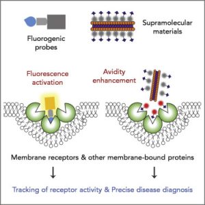
Studying the dynamics of cell surface receptors is critical to understanding how cells sense and respond to changing environments. Therefore, elucidating the mechanisms by which signals are perceived and communicated to the cell is necessary to understand immunity, development, and stress. Challenges in testing membrane-bound protein interactions include their dynamic nature, their abundance, and the complex dual environment (lipid/soluble) in which they reside. Co-immunoprecipitation (Co-IP) of tagged membrane proteins is a widely used approach to test protein-protein interaction in vivo.
In this protocol, we present a method to perform Co-IP using membrane-bound receptors proteins enriched in isolated microsomal fractions. Different variations of this protocol are highlighted, including recommendations and troubleshooting guides to optimize your application. This Co-IP protocol has been developed to test the interaction of receptor-like kinases, their interacting partners, and peptide ligands in stable lines of Arabidopsis thaliana, but can be modified to test interactions in transiently expressed proteins in tobacco and potentially other plants. models, or scaled for large-scale protein-protein interactions in the membrane.
Membrane-bound receptors and the second messenger concept
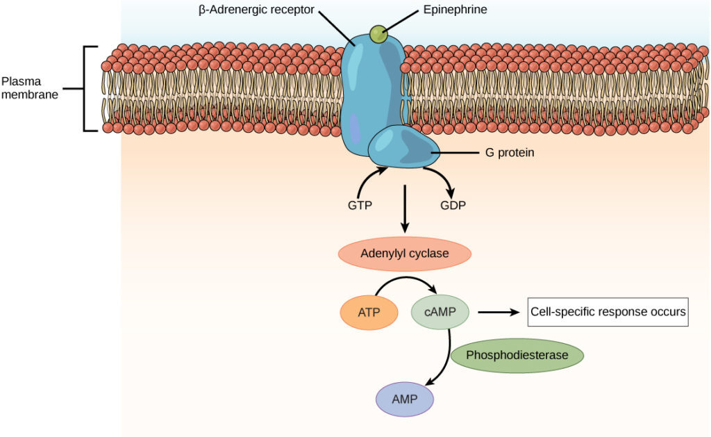
Many hormones, such as most amino acid derivatives and peptide hormones that are too large or too polar to cross cell membranes, bind to receptor sites on the surface of target cell membranes. The hormone and receptor form a complex that triggers a cascade of molecular events within a cell. The hormone thus behaves as a first messenger causing the activation of a second messenger system in the cytoplasm. At least six different molecules have been identified as second messengers. Each acts through a specific kinase, which causes the activation or inactivation of rate-limiting enzymes that change the direction and rate of cytoplasmic processes. Since many second messenger molecules are activated after a single hormone molecule has been bound, the message is amplified, perhaps thousands of times.
Second messenger systems known to be involved in hormonal actions are cyclic AMP (cAMP), cyclic GMP (cGMP), Ca++/calmodulin, inositol trisphosphate (IP3), and diacylglycerol (DAG). Cyclic AMP was the first to be investigated and has been shown to mediate the actions of many peptide hormones, including parathyroid hormone, glucagon, adrenocorticotropic hormone (ACTH), thyrotropic hormone (TSH), melanophore-stimulating hormone ( MSH) and vasopressin. It also mediates the action of epinephrine (also called adrenaline), an amino acid derivative.
Types of Receptors
1. Internal receptors
Internal receptors, also known as intracellular or cytoplasmic receptors, are found in the cytoplasm of the cell and respond to hydrophobic ligand molecules that can travel across the plasma membrane. Once inside the cell, many of these molecules bind to proteins that act as regulators of mRNA synthesis. Remember that mRNA carries genetic information from the DNA in a cell’s nucleus to the ribosome, where protein is assembled.
When the ligand binds to the internal receptor, it triggers a shape change that exposes a DNA-binding site on the receptor protein. The ligand-receptor complex moves into the nucleus then bind to specific regions of the DNA and promote the production of mRNA from specific genes. Internal receptors can directly influence gene expression (how much specific protein is produced from a gene) without having to pass the signal to other receptors or messengers.
2. Cell Surface Receptors
Cell surface receptors, also known as transmembrane receptors, are proteins that are found attached to the cell membrane. These receptors bind to external ligand molecules (ligands that do not travel through the cell membrane). This type of receptor crosses the plasma membrane and performs signal transduction, in which an extracellular signal is converted into an intercellular signal. Ligands that interact with cell surface receptors do not have to enter the cell they affect. Cell surface receptors are also called cell-specific proteins or markers because they are specific to individual cell types.
Each cell surface receptor has three major components: an external ligand-binding domain, a membrane-spanning hydrophobic region, and an intracellular domain within the cell. The size and extent of each of these domains vary widely, depending on the type of receptor. Cell surface receptors are involved in most signalling in multicellular organisms. There are three general categories of cell surface receptors: ion channel-linked receptors, G protein-linked receptors, and enzyme-linked receptors.
3. Receptors linked to ion channels
Ion channel-linked receptors bind a ligand and open a channel through the membrane that allows the passage of specific ions. To form a channel, this type of cell-surface receptor has an extensive membrane-spanning region. When a ligand binds to the extracellular region of the channel, a conformational change occurs in the structure of the proteins that allow the passage of ions such as sodium, calcium, magnesium, and hydrogen.
4. G protein-coupled receptors
G protein-coupled receptors bind a ligand and activate a membrane protein called a G protein. The activated G protein then interacts with an ion channel or enzyme in the membrane. Before the ligand binds, the inactive G protein can bind to a site on a specific receptor. Once the G protein binds to the receptor, the G protein changes shape, becomes active, and splits into two different subunits. One or both of these subunits can activate other proteins as a result.
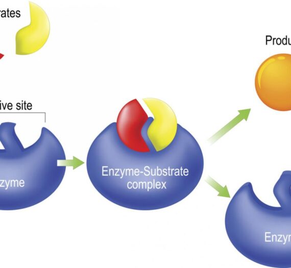
5. Enzyme-linked receptors
Enzyme-linked receptors are cell surface receptors with intracellular domains that are associated with an enzyme. In some cases, the intracellular domain of the receptor itself is an enzyme. Other enzyme-linked receptors have a small intracellular domain that directly interacts with an enzyme. When a ligand binds to the extracellular domain, a signal is transferred across the membrane, activating the enzyme. Activation of the enzyme sets off a chain of events within the cell that eventually leads to a response.